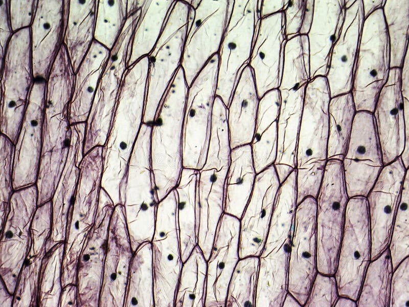
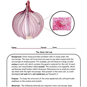
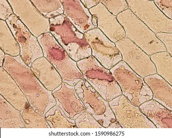
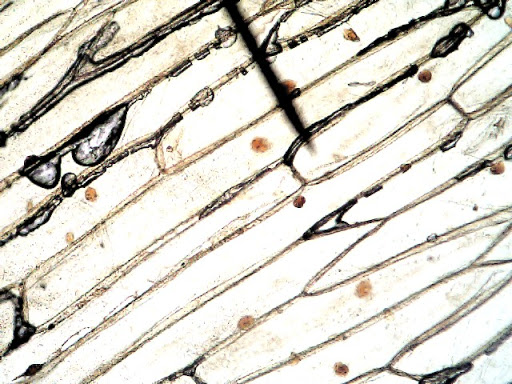
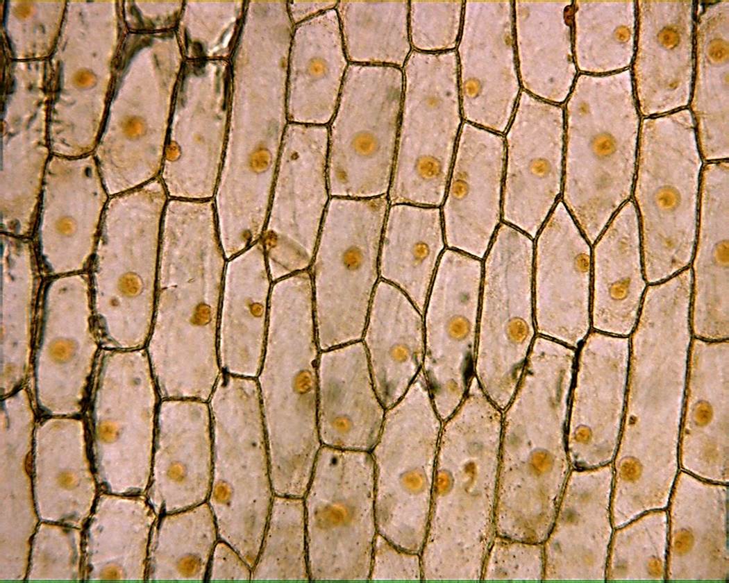
40 onion cells under microscope with labels
Onion cell under microscope 4x - raa.svleasing.pl Onion cell under microscope 4x The observation she most likely makes is: the onion cells have a cell wall, and the cheek cells do not The observation she most likely makes is: the onion cells have a cell wall, and the cheek cells do not. Materials and Equipment: Microscope Slides (2) Cover Slips (2) Eye Droppers Beaker Toothpick Water *Red dye. Examine a variety of cells with the compound ... Onion cells under the microscope: 40X - 100X - 400X - YouTube under the #microscope: 40X - 100X - 400X
Onion Cells Under Microscope With Labels - Realtec Find and download Onion Cells Under Microscope With Labels image, wallpaper and background for your Iphone, Android or PC Desktop. Realtec have about 34 image published on this page. …

Onion cells under microscope with labels
Observing Cork Cells Under The Microscope » Microscope Club Place the cork dust on the microscope slide with a drop of water, then add another water droplet on top of the cork sample. Cover the prepared slide with a cover slip. Method 2 Alternatively, slice thin cork slices, making sure that ample light can pass through the slice, allowing you to see the cell layout and the individual cells. Under the Micrsocope: Onion Cell (100x - 400x) - YouTube 22/07/2013 · In this "experiment" we will see onion cells under the microscope.For the experiment you will only need onion, dropper and the microscope (container and tool... Onion Root Cells Dividing By Mitosis Under A Light Microscope ... - iStock Download this Onion Root Cells Dividing By Mitosis Under A Light Microscope At 100x Magnification Cells Visible In Prophase Metaphase Anaphase And Telophase photo now. And search more of iStock's library of royalty-free stock images that features Anaphase photos available for quick and easy download.
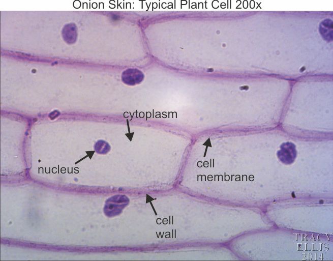
Onion cells under microscope with labels. The following diagram shows cells of onion peel label class ... - Vedantu Grade 11 Cell Answer The following diagram shows cells of onion peel, label the cell wall, nucleus and cytoplasm. The following diagram shows human cheek cells, label the parts as observed by you. Answer Verified 144k + views Hint: The diagrams mentioned above are the internal structure of an onion peel and human cheek cells. Microscope Cell Lab: Cheek, Onion, Zebrina The onion epidermis cell is the only cell that has a cell wall. In addition, it is the only cell that has a chloroplast, where photosynthesis can happen. The cheek epithelium cell is the only one that has centrioles, the barrel-shaped organelle that is responsible for helping organize chromosomes during cell division. DOC Plant and Animal Cells Microscope Lab - hillsboro.k12.oh.us Make a drawing of one onion cell, labeling all of its parts as you observe them. (At minimum you should observe the nucleus, cell wall, and cytoplasm.) Cheek cells 1. To view cheek cells, gently scrape the inside lining of your cheek with a toothpick. DO NOT GOUGE THE INSIDE OF YOUR CHEEK! (We will observe blood cells in a future lab!!) 2. Onion Cells Under a Microscope Add a drop of iodine solution on the onion membrane (or methylene blue) Gently lay a microscopic cover slip on the membrane and press it down gently using a needle to remove air bubbles. Touch a blotting paper on one side of the slide to drain excess iodine/water solution, Place the slide on the microscope stage under low power to observe.
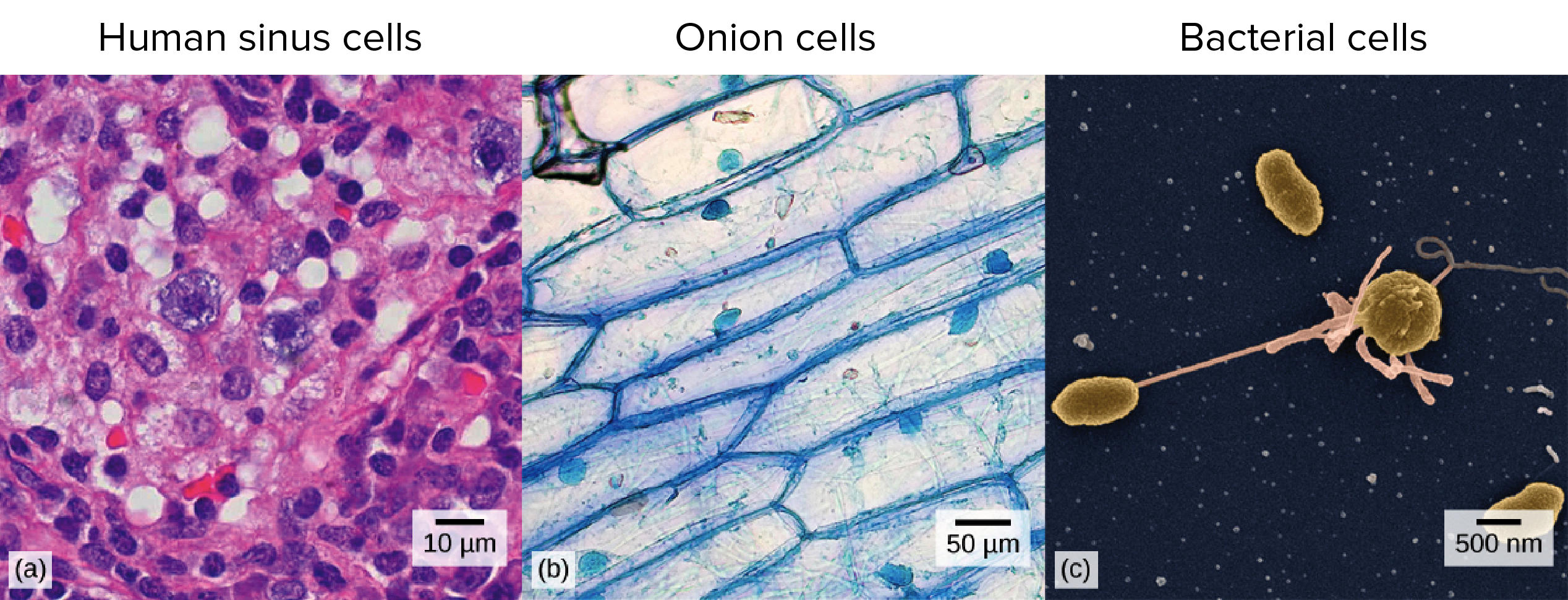
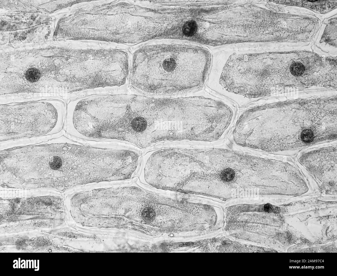
Onion Skin Epidermis Sample under microscope 4x,10x Magnification A sample on an onion skin epidermis diyed in blue for visibility, viewd under the microscope at 4x and 10x magnification.microscope:Biolux model :AL [Solved] What organelles are in an onion cell? | 9to5Science To answer your question, onion cells (you usually use epithelial cells for this experiment) are 'normal' cells with all of the 'normal' organelles: nucleus, cytoplasm, cell wall and membrane, mitochondria, ribosomes, rough and smooth endoplasmic reticulum, centrioles, Golgi body and vacuoles. Lesson 3: Onion Dissection & “Look at the Plant Cells” Preparing onion cells slide for a microscope. Peel the brown skin away from the outside of the onion. Take one layer of the onion flesh and carefully cut out a piece. On the inside of this piece is a thin sheet of the membrane. Use … Onion Cells Microscope Stock Photos and Images - Alamy RM2AM97C0-Onion skin cells under the microscope, horizontal field of view is about 0.61 mm RFHWA476-Onion epidermis with large cells under light microscope. Clear epidermal cells of an onion, Allium cepa, in a single layer. RM2DF6FFJ-Onion epidermis (Allium cepa) showing cells and nucleus. Optical microscope X200.
Collins - Concise Revision Course For CSEC Biology PDF 5 Cells The cell is the basic structural and functional unit of living organisms. Some organisms are unicellular, being composed of a single cell; others are multicellular, being composed of many cells. Cells are so small that they can only be seen with a microscope and not with the naked eye. Images, Stock Photos & Vectors | Shutterstock Jun 30, 2022 · Find stock images in HD and millions of other royalty-free stock photos, illustrations and vectors in the Shutterstock collection. Thousands of new, high-quality pictures added every day. K To 12 Science Grade 7 Learners Material - Module | PDF ... draw onion cells as seen through the light microscope; and 6. explain the role of microscopes in cell study. Materials Needed dropper tissue paper cover slip iodine solution glass slide light microscope onion bulb scale forceps or tweezers scalpel or sharp blade 50-mL beaker with tap water Procedure 1. Prepare the onion scale by following steps ... Observing Onion Cells Under The Microscope » … Afterwards, carefully mount the prepared and stained onion cell slide onto the microscope stage. Make sure that the cover slip is perfectly aligned with the microscope slide, and that any excess stain has been wiped off. Secure the slide on the stage using the stage clips.
Onion Cell Diagram Labeled Pdf (PDF) - thesource2.metro Set your multimeter to measure current in the 20 mA range (the dial setting labeled "20m" on the right). Plug the multimeter's black probe into the port labeled COM. Plug the multimeter's red probe into the port labeled VΩmA. Use a red alligator clip lead to connect the multimeter's red probe to the positive (+) terminal of the 9 V battery.
Plant Cell Under Microscope Drawing When viewing onion cells under a microscope a few drops of a certain solution are added to stain the cells and show these cells more clearly. Ii From the solutions given in brackets water strong sugar solution 1 salt solution name the solution into which 1. The Figure Below Is A Fine Structure Of A Generalized Animal Cell.
Publications – Pradeep Research Group 2 days ago · Facile crystallization of ice I h via formaldehyde hydrate in ultrahigh vacuum under cryogenic conditions, Jyotirmoy Ghosh, Gaurav Vishwakarma, Subhadip Das, and Thalappil Pradeep, J. Phys. Chem. C, 125 (2021) 4532–4539 (DOI: 10.1021/acs.jpcc.0c10367). PDF File Supporting Information
The Cell - ScienceQuiz.net The diagram shows a group of onion cells. The parts labelled A, B and C respectively are ... The diagram shows a plant cell as seen under a microscope. Two of the ...
Onion Peels Observed Under the Microscope | Confirmation Point Onion Peels Observed Under the Microscope Cells present in onion peel can be observed under microscope. For this onion peels are first isolated. For this experiment outer most scale of the onion is removed and is cut into four equal halves. It is a monocot plant. Then with the help of a pairs of forcep the scale of onion is peeled out.
Microscopy Practical (Onion Cells) | Teaching Resources 20/01/2022 · pptx, 12.37 MB. pdf, 223.81 KB. docx, 1.09 MB. Presentation and practical handout for observing onion cells under a light microscope for teaching and revision. A step by step visual guide for all abilities. Can be used as a …
Onion Cells Under a Microscope (100x-2500x) - YouTube 24/02/2021 · In this video you will see onion cells under a microscope (100x-2500x) as is, without any coloring. To observe the onion cells the thin membrane is used. It...
Onion Plant Cell Under Microscope Labeled - Ismael Dauila Explore diffusion/osmosis by looking at onion cells under the microscope. It is used for treating a parasite disease called ich (ichthyophthirius multifiliis; Label the cell wall and chloroplasts. Students will observe plant cells using a light microscope.
Onion Cell Lab Report.docx - Onion Cell Lab Report By station, remove the single layer of epidermal cells from inner side of the scale leaf. 3(Place the single layer of onion on a glass slide. 4(Place a drop of iodine stain on your onion tissue. 5(Put the cover slip on the stained tissue and gently tap out any air bubbles. 6(Observe the cells under the microscope and see you results.
(PDF) Cambridge International AS and A Level Biology ... • Enzymes are globular proteins; the basic building blocks of enzymes are amino acids • Their manufacture is controlled by nucleus • Are needed only in small amounts • Enzymes are biological catalysts; they control the rate of a reaction, but are chemically unchanged at the end of the reaction • Enzymes are specific, they affect only ...
The Cell Structure of an Onion | Sciencing Cell Walls Give Structure. Cell walls in plants are rigid, compared to other organisms. The cellulose present in the cell walls forms clearly defined tiles. In onion cells the tiles look very similar to rectangular bricks laid in offset runs. The rigid walls combined with water pressure within a cell provide strength and rigidity, giving plants ...
Animal Cell Mitosis Under Microscope - Casey Sillman The division of the cell in two (cytokinesis) occurs chromosomes decondense (no longer visible under light microscope). In cell biology, mitosis (/maɪˈtoʊsɪs/) is a part of the cell cycle in which replicated chromosomes are separated into two new nuclei. Plant cells do not have centrioles like animal cells, just centrosomes.
The Biology Project The Biology Project, an interactive online resource for learning biology developed at The University of Arizona. The Biology Project is fun, richly illustrated, and tested on 1000s of students.
Plant Cell Under Microscope 40X / Plant Cells Under ... - Blogger Assignment 6 Page 2 from projects.ncsu.edu Label the cell wall, cytoplasm (cyto = cell). Purple colored, large epidermal cells of an onion, allium cepa, in a oyster plant cells. ... This section on microscopy is meant as an introduction as learners will need to be able to use these same onion cells were viewed under a microscope which had not ...
Cheek Cells Under a Microscope - Requirements/Preparation/Staining smear the cotton swab on to the center (part containing the saline drop) of the clean slide for about 4 seconds to get the cells on to the center of the slide add a drop of methylene blue solution on to the smear and gently place a cover slip on top (to cover the stain and the cells)
2022. 7. 23. · Search: Human Cheek Label the cell wall, cytoplasm, and the pigmented organelle structures (but you must label it with their real name 1cm2 is sufficient Sketch - diagram of onion cells as seen under a microscope This makes a root tip an excellent tissue to study the stages of cell division Obtain a slide of onion root cells Obtain a slide of onion root cells. 2022.
What organelles are in an onion cell? - Biology Stack Exchange You cannot see most of these as they appear translucent as well as being too small to see under the light microscope. You need an electron microscope to view these. Note: chloroplasts are not present in an onion cell as it is not a photosynthesising cell. This is a typical onion cell slide with labels:
Onion Root Tip Mitosis - Stages, Experiment and Results - MicroscopeMaster · Place a cap/lid onto the vial (ensure that the cap/lid has a pinprick hole) and place the vial in the water bath (at 55 degrees C) for about 5 minutes - This enhances the staining process · Using the forceps, remove the root tips from the vial of stain and place them onto a clean microscope glass slide
Onion Skin Cells - Investigation Observe the onion tissue under the microscope at 4x, 10x and 40x with lots of light (open diaphragm). Then slowly close the diaphragm while observing the image to find the best light for seeing cellular details. 6. Draw a section of onion skin cells at 10x magnification. Then switch to 40x and draw one cell and label it.
Leaf Structure Under the Microscope Allow the nail polish about four hours to dry. Using a pair of tweezers, peel off a film (thin skin) from the surface of the leaf. Gently place the film onto a microscope slide and cover with a cover slip. Start with low power and increase to 100x (frequency of stoma can be counted at 100x) Record your observations.
Plant Cell Under Microscope Labeled 40X - Sadie Bermingham Cells and viewing them under the microscope. A small square of a red onion skin (membrane) was observed under a microscope at high power (x40) magnification. (iv) describe how you applied the stain. They must draw and label the nucleus, cell membrane set up your microscope, place the onion root slide on the stage and focus on low (40x) power.
Onion Root Cells Dividing By Mitosis Under A Light Microscope ... - iStock Download this Onion Root Cells Dividing By Mitosis Under A Light Microscope At 100x Magnification Cells Visible In Prophase Metaphase Anaphase And Telophase photo now. And search more of iStock's library of royalty-free stock images that features Anaphase photos available for quick and easy download.
Under the Micrsocope: Onion Cell (100x - 400x) - YouTube 22/07/2013 · In this "experiment" we will see onion cells under the microscope.For the experiment you will only need onion, dropper and the microscope (container and tool...
Observing Cork Cells Under The Microscope » Microscope Club Place the cork dust on the microscope slide with a drop of water, then add another water droplet on top of the cork sample. Cover the prepared slide with a cover slip. Method 2 Alternatively, slice thin cork slices, making sure that ample light can pass through the slice, allowing you to see the cell layout and the individual cells.













Post a Comment for "40 onion cells under microscope with labels"